
Skull Base Surgery
Clinical Interests Last updated: Author: Yücel Doğruel, MD, Neurosurgeon
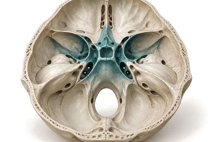
The skull base is the anatomical region beneath the brain through which the major cerebral vessels and the nerves serving important functions such as vision, hearing, facial movement and swallowing pass. Tumours that develop here call for a special surgical assessment, not only because of their size but because of their relationship to the delicate structures around them.
The principal aim of skull base surgery is to control the disease while preserving neurological function as far as possible. The treatment decision depends on the type of tumour, its location, its growth, the symptoms it causes and its relationship to the surrounding anatomical structures. Not every skull base tumour requires surgery.
Which diseases?
Skull base meningiomas
Meningiomas, which arise from the membranes covering the brain, can occur at various sites, including the sphenoid wing, the anterior skull base, the tuberculum sellae, the petroclival region and the tentorium. The position of the tumour and its relationship to the optic nerves and the major vessels are important in treatment planning.
Cerebellopontine angle tumours
Lesions such as vestibular schwannoma (acoustic neuroma), meningioma and epidermoid cyst can develop in the region between the cerebellum and the brainstem (the cerebellopontine angle). These tumours may affect the nerves serving hearing, balance and facial movement.
Cranial nerve tumours
Tumours arising from the cranial nerves, trigeminal schwannomas above all, may extend into different compartments along the course of the nerve through the skull base. In the surgical assessment, the relationship of the tumour to the nerve fibres, the brainstem and the surrounding vessels is decisive.
Craniopharyngiomas and suprasellar tumours
Tumours located above the pituitary gland, close to the visual pathways and the hypothalamus. They may cause visual disturbances, hormonal problems and, in some cases, obstruction of the circulation of cerebrospinal fluid. Preserving the visual pathways and the function of the pituitary gland and hypothalamus is especially important in the treatment plan.
Orbital and anterior skull base tumours
Tumours arising from the orbit (eye socket) and the neighbouring skull base structures may be closely related to the optic nerve, the nerves controlling eye movement and the surrounding vessels. The surgical approach is chosen according to the position of the tumour within the orbit and its relationship to the skull base.
What symptoms can skull base tumours cause?
Symptoms vary with the region in which the tumour lies. Reduced vision or double vision, hearing loss, tinnitus, imbalance, numbness or pain in the face, weakness of facial movement and headache may occur.
Some tumours may also cause difficulty in swallowing, a change in the voice or hormonal disturbances. Small, slow-growing tumours, on the other hand, may remain without symptoms for a long time.
These complaints are not specific to skull base tumours. The diagnosis is assessed by neurological examination and appropriate imaging studies.
When is surgery needed for a skull base tumour?
The decision to operate is not based on the size of the tumour alone. Surgical treatment may be considered for tumours that are growing, that compress the brainstem or nerve structures, or that affect vision or other neurological functions.
For some small, asymptomatic or slow-growing tumours, however, regular MRI follow-up may be appropriate. Depending on the features of the tumour, stereotactic radiosurgery or radiotherapy are also among the treatment options.
When surgery is planned, the goal in suitable cases is complete removal of the tumour. Where the tumour is firmly adherent to an important nerve, vessel or the brainstem, however, a limited resection followed by observation or additional treatment may be preferred in order to preserve neurological function.
Diagnosis and surgical planning
Contrast-enhanced MRI of the brain is the principal imaging method for assessing skull base lesions. Thin-slice CT may be used where the bony structures need detailed examination, and CT angiography or other vascular imaging where the relationship to the vessels has to be defined.
In patients who need them, additional studies such as hearing tests, visual field examination, ophthalmological examination and hormonal assessment support the treatment decision.
In surgical planning, the relationship of the tumour to the vessels, the cranial nerves, the brainstem and the surrounding bony structures is assessed as a whole. In selected cases, patient-specific three-dimensional models are built so that the surgical corridor, the position of the craniotomy and the direction of the approach can be examined before the operation.
Knowledge of microsurgical neuroanatomy plays a fundamental role in choosing, among the various surgical routes, the approach suited to the lesion. Where required, intraoperative neuromonitoring is used during surgery to monitor the function of the cranial nerves.
Surgical treatments
Microsurgical skull base approaches
Depending on the location of the tumour, different microsurgical approaches such as the pterional, retrosigmoid, supracerebellar or interhemispheric approach may be used to reach skull base tumours.
The choice of approach rests on identifying the most suitable anatomical corridor to the tumour. For different tumours in the same region, different surgical routes may be preferred according to their relationship to the vessels and nerves.
Endoscope-assisted and other surgical approaches
In some skull base lesions the endoscope can help in assessing areas that lie outside the direct line of sight of the microscope. In suitable anatomical locations, endonasal and other specialised skull base approaches are also among the treatment options.
In orbital tumours, different surgical corridors, including transmaxillary approaches in selected cases, may be considered according to the position of the lesion.
Partial removal and radiosurgery
In some patients, where complete removal of the tumour would carry a serious neurological risk, reducing the volume of the tumour and following the remainder with imaging or treating it with radiosurgery may be appropriate.
This approach is considered in particular for certain skull base tumours in which preserving the cranial nerves is the priority.
The postoperative period and follow-up
Postoperative follow-up is planned according to the type of tumour, the extent of the surgical removal and the patient’s neurological condition.
MRI is used to assess the surgical field and any remaining tumour tissue. Where required, additional assessments of vision, hearing, facial movement, swallowing and hormonal function are carried out.
Depending on the results of the pathological examination and the features of the tumour, regular follow-up, radiosurgery or radiotherapy may be needed.
Frequently asked questions
Do skull base tumours always need surgery?
No. Some small tumours that cause no symptoms can be followed with regular MRI. The treatment decision depends on the type of tumour, its growth, its location and the symptoms it causes.
Can a skull base tumour be removed completely?
Some tumours can be removed completely. In tumours firmly adherent to important nerves and vessels, however, leaving part of the tumour in place may be the better option in order to preserve neurological function.
Can hearing or facial movement be affected by skull base surgery?
Yes. In cerebellopontine angle tumours in particular, there is a risk to the nerves serving hearing and facial movement. This risk varies with the size and location of the tumour and its relationship to the nerves.
Can skull base tumours be operated on through the nose?
Some midline skull base tumours can be reached by endoscopic endonasal approaches. Not every tumour is suitable for this method, however. The choice between open microsurgical and endoscopic approaches is made according to the anatomical features of the tumour.
Might radiosurgery be needed after surgery?
It may be needed in some patients. Radiosurgery or radiotherapy may be considered, particularly where part of the tumour has been left in place or where there is a risk of growth.
Information needed for the first assessment
It is important that the current contrast-enhanced brain MRI can be reviewed, in DICOM format if possible, together with any previous studies.
Depending on the location of the disease, the results of hearing tests, visual field examinations or hormonal assessment may be useful. If surgery or radiosurgery has been performed before, the relevant reports also help in understanding the treatment history.
For referring physicians
In skull base cases, thin-slice contrast-enhanced MRI is usually sufficient for planning; the need for bone-window CT and CT angiography is decided case by case. A DICOM copy of the images together with details of previous surgery and radiosurgery, hearing test results and the findings of the cranial nerve examination make the assessment easier. Professional correspondence: yucel@yuceldogruel.com
Related publications
The information on this page is for general purposes only. Diagnosis and treatment decisions are made on the basis of each patient’s individual assessment.